Fibrosis is driven by the excessive production and deposition of collagen and other extracellular matrix proteins in tissues and organs. The increase in tissue stiffness or scarring associated with fibrosis impacts the normal functioning of tissues. Fibrosis-associated diseases include idiopathic pulmonary fibrosis (IPF), nonalcoholic steatohepatitis (NASH), cirrhosis (LF), hepatitis, chronic kidney disease (CKD), myocardial infarction, heart failure, celiac disease, scleroderma, diabetes and more.
Aragen has a portfolio of in vivo, in vitro and ex vivo fibrosis models and assays as well as world class expertise to support clients in the development of anti-fibrotic therapeutic candidates. In hundreds of studies, over the course of more than a decade, we have evaluated the efficacy of test compounds spanning several modalities (small molecules, biologics, RNA therapeutics, exosomes etc.) on a variety of assay platforms, ultimately resulting in successful IND submissions for our clients. Aragen’s diet-induced, chemical-induced, and surgical models of fibrosis, coupled with an extensive evaluation of disease parameters can help drive research efforts towards finding novel therapies.
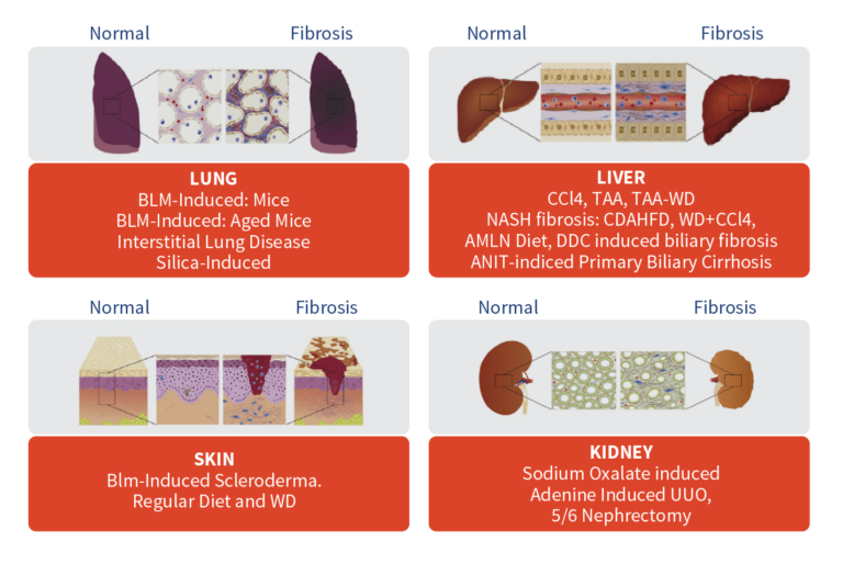
Abbreviations: Blm- Bleomycin; CCl4-Carbon Tetrachloride; TAA-Thioacetamide; WD- Western Diet; UUO- unilateral ureteral obstruction; NASH- nonalcoholic steatohepatitis; CDAHFD- L-amino acid-defined, high-fat diet; AMLN- amylin liver non-alcoholic steatohepatitis; ANIT- Alpha-naphthyl isothiocyanate.
In Vivo Preclinical Models of Fibrosis
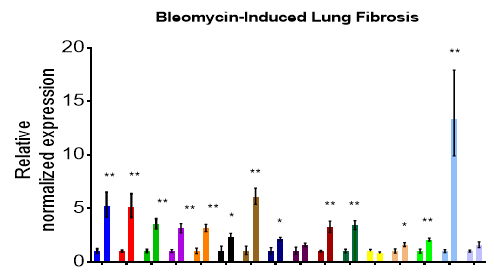
Lung Fibrosis Model in C57BL/6 Mice
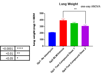
The most common preclinical animal model for idiopathic pulmonary fibrosis (IPF) is the rodent model of bleomycin-induced pulmonary fibrosis. Bleomycin induction causes inflammatory and fibrotic reactions within a short period of time in rodent lungs.
Bleomycin-induced lung fibrosis followed by treatment through a range of administration routes (PO, IP, IV, SC, IM, ID, intranasal or sublingual). Ex vivo analysis can include a variety of readouts.
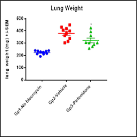
Method- Bleomycin-Induced Pulmonary Fibrosis (IPF) Model in Mice
- Study animals: C57BL/6
- Fibrosis induction: Clinical grade Bleomycin-instilled via oropharyngeal route or osmotic pump
- Option of test article administration: PO, IP, IV, IM, SC, nebulization, and osmotic pumps
- Treatment regimen: Therapeutic or Prophylactic
- Positive control (standard of care): Pirfenidone, Nintedanib, ALK 5i
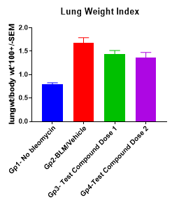
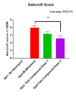
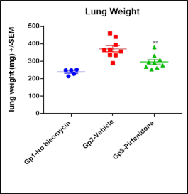
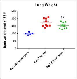
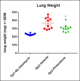
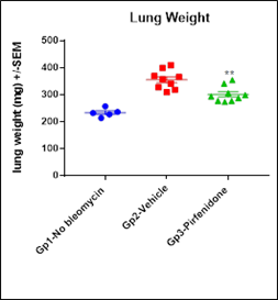
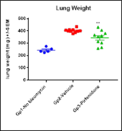
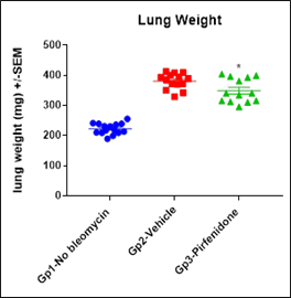
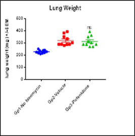
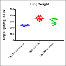
Treatment groups
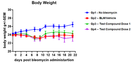
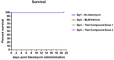
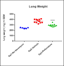
- Gp1- No Bleomycin
- Gp2-Bleomycin/Vehicle
- Gp3-Pirfenidone
Key Readouts Standard
- Body weight
- Survival
- Lung weight
- Leukocyte count in bronchoalveolar lavage (BAL)
- Lung Histology Analysis
Fibrosis Measurements
- Lung hydroxyproline
- Serum/BAL soluble mediators
- Lung FibroPanelTM Gene expression
- Lung Histology Analysis
- Differentials from BAL cells
Lung Function Measurements
- flexiVentTM
- Hypoxia related parameters
- Whole-body plethysmography
Lung Function Analyses
- flexiVentTM analysis demonstrates that bleomycin instillation increases resistance and elastance while decreasing compliance.
- Enhanced Respiratory pause (Penh) is increased by Bleomycin.
- Pirfenidone improves lung function measurements.


Highly reproducible and consistent fibrotic induction in lungs allows for robust evaluation of candidate drugs.
Lung Fibrosis Model – Aged Mice
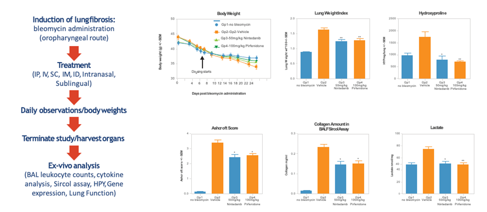
Progress has been made in developing rodent models that demonstrate an age-dependent increase in fibrosis, more closely mimicking the pathogenesis of human IPF. In this model, lung fibrosis is induced by bleomycin followed by treatment through a range of administration routes (IP, IV, SC, IM, ID, intranasal or sublingual). Ex vivo analysis can include a variety of readouts.
Method
- Gp1-no bleomycin
- Gp2-Gp2-Vehicle
- Gp3-50mg/kg Nintedanib
- Gp4-100mg/kg Pirfenidone
Key Readouts- Ex-vivo analysis
- BAL leukocyte counts
- Cytokine analysis
- Sircol assay
- Hydroxyproline
- Gene expression
- Lung function
- Histology & Digital Pathology
- Immunohistochemistry (IHC)
Silica-Induced Lung Fibrosis in Mice
Aragen has developed a silica-induced pulmonary fibrosis model in mice. Mice are instilled with crystalline silica or saline via oropharyngeal route. Silica-instillation causes persistent inflammation of alveoli leading to inflammatory and fibrotic processes not resolving with time. This model of non-resolving lung fibrosis provides quantitative assessment of disease progression over a longer period, essential towards evaluation of fibrotic disease burden and antifibrotic therapy evaluation in preclinical studies. Treatment, as usual, can be through a range of administration routes (IP, IV, SC, IM, ID, intranasal or sublingual). Ex vivo analysis can include a variety of readouts.
The gene expression profile presents a profile that is different compared to bleomycin induced lung fibrosis model suggesting the activation of different pathways in both models.
Method
- Gp1-no silica
- Gp2-Silica
Key Readouts- Ex-vivo analysis
- BAL leukocyte counts
- Cytokine analysis
- Sircol assay
- Hydroxyproline
- Gene expression
- Lung Function
- Histology & Digital Pathology
- IHC
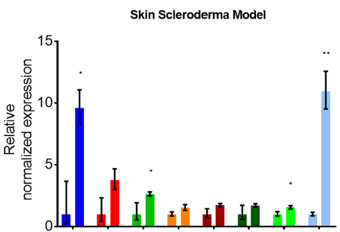
Systemic Sclerosis/Scleroderma Mouse Model
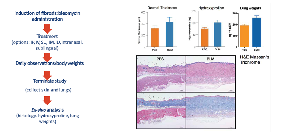
Bleomycin-induced dermal fibrosis is a well-characterized experimental model used to evaluate the potential role of genes or treatments in the early inflammatory phase and in the subsequent fibrosis process typical for scleroderma. In this model, after bleomycin administration and treatment administered through a range of routes (IP, IV, SC, IM, ID, intranasal, inhalation), ex vivo analysis of skin or lungs is performed to evaluate the efficacy of the test compound.
Method
- Gp1-PBS
- Gp2-BLM
Key Readouts- Ex-vivo analysis
- Histology & Digital Pathology
- IHC
- hydroxyproline
- lung weights
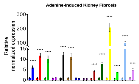
Renal Interstitial Fibrosis Mouse Model
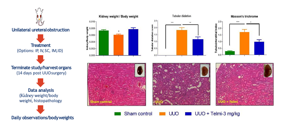
The Unilateral Ureteral Obstruction (UUO) model is used to cause renal fibrosis in mice, where the primary feature of UUO is tubular injury because of obstructed urine flow. UUO in rodents is believed to mimic human chronic obstructive nephropathy in an accelerated manner.
After obstruction, treatment may be administered through a range of routes (IP, IV, SC, IM, ID, intranasal, sublingual), and ex-vivo analysis of the kidneys were performed.
This model is routinely used for preclinical efficacy testing of NCEs for treatment of renal nephropathy/fibrosis.
Method
- Gp1-Sham
- Gp2-UOO
- Gp3-UOO + Telmisartan (Telmi-3 mg/Kg)
Key Readouts- Ex-vivo analysis
- Body/ Kidney Weight
- Tubular Dilation
- Gene Expression
- Cytokine Analysis
- Histology & Digital Pathology
- IHC
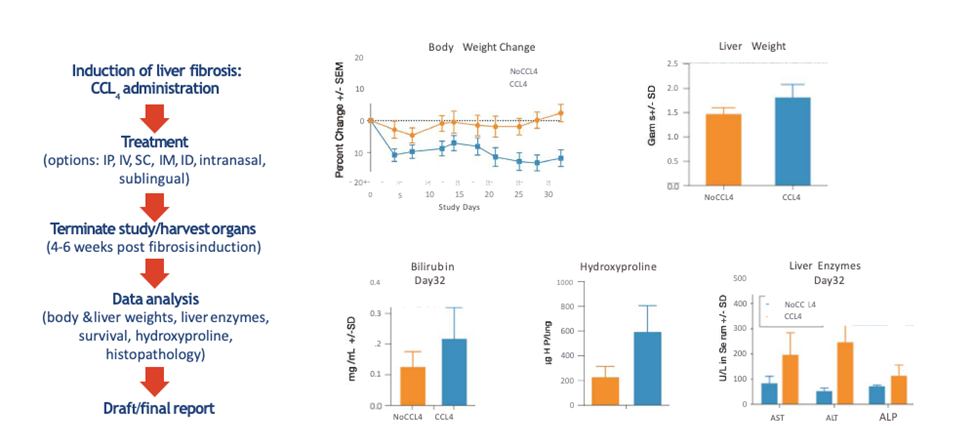
CCl4 Induced Liver Fibrosis Model in Rodents
Carbon tetrachloride (CCl4) has been widely used to experimentally induce liver injury in rodents. After a period of dosing with CCl4, treatment is administered through a range of routes (IP, IV, SC, IM, ID, intranasal, inhalation). Key readouts include ex vivo analysis of the liver.
Method
- Gp1-No CCl4
- Gp2- CCl4: Mineral oil
Key Readouts
- Body Weight
- Ex vivo Analyses (liver weights, liver enzymes, clinical chemistry, histology, hydroxyproline,)
- Fibro PaneI (gene expression analysis)
- Gene Expression
- Protein Expression
- Histology & Digital Pathology
- IHC
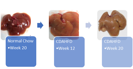
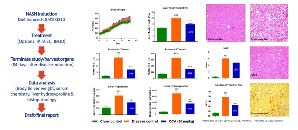
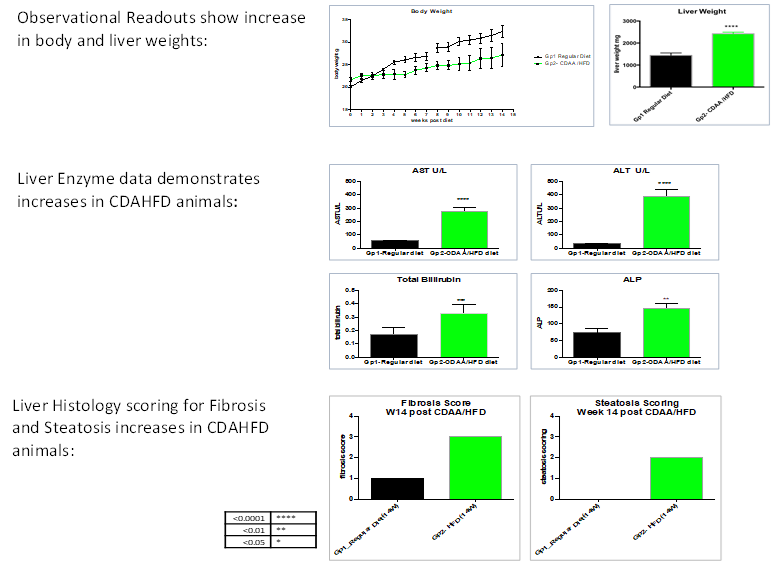
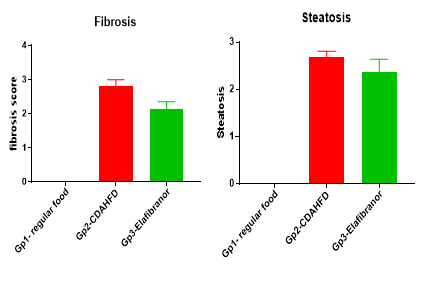
Diet-Induced Non-Alcoholic Steatohepatitis (NASH) in Mice
- Study animals: C57BL/6
- Disease induction: Choline-deficient, L-amino acid-defined, high-fat diet (CDAHFD) consisting of 60 kcal% fat and 0.1% methionine.
- Option of test article administration: PO, IP, IV, IM, SC, nebulization, and osmotic pumps
- Treatment regimen: Therapeutic or Prophylactic
- Model developed as described by Matsumoto et al., 2013 Int. J. Exp. Pathol.
- Rodent Diet:40 kcal% Fat (Mostly Palm Oil), 20 kcal% Fructose and 2% Cholesterol (Source Research Diets, D09100310).
Method
- Gp1-CHOW control
- Gp2-CDAHFD
Key Readouts
- Body Weight
- Daily Activity
- Survival
- Serum Liver Enzymes
- Liver Histology: H&E and Picrosirius Red (PSR) Staining
- Hepatic Hydroxyproline Content
- Serum/Plasma Biomarkers
- Liver FibroPanelTM Gene Expression
- Abdominal Fat Collection
- Liver Histology: H&E and Picrosirius Red (PSR) Staining
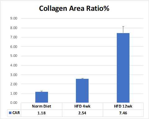
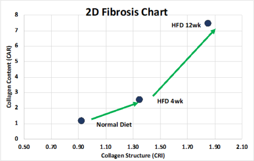
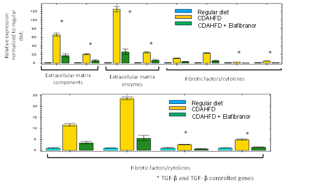
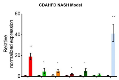
Characterization of CDAHFD, NASH-Fibrosis model
Representative Second Harmonic Generation images from Normal Diet & CDAHFD control group animals:
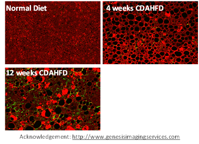
Analysis of Collagen ratio and structure in animals post normal and ADHDFD diets feeds.

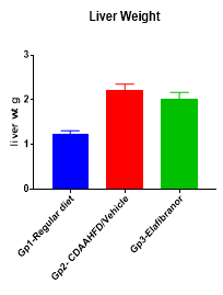
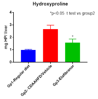
FibroPanelTM gene expression data demonstrates the effects of Elafibranor treatment on CDAHFD animals:
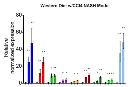
Non-Alcoholic Steatohepatitis Model: Western Diet + CCl4 Induced Method
- Study animals: C57BL/6
- Disease induction: Western diet with weekly administration of Carbon Tetrachloride (CCl4)
- Option of test article administration: PO, IP, IV, IM, SC, nebulization, and osmotic pumps
- Treatment regimen: Therapeutic or Prophylactic
- References: Model adapted from Tsuchida et al., 2018 J Hepatol.
Key Readouts
- Body weight
- Daily activity
- Survival
- Serum liver enzymes
- Liver histology:
- Hydroxyproline
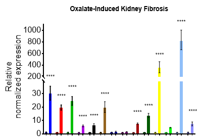
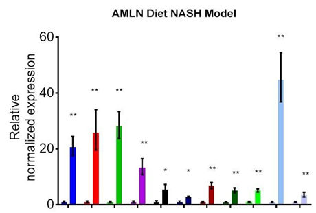
Linking Multiple Preclinical Fibrosis Models through Gene Expression
Multiple preclinical fibrosis models can be linked through gene expression analysis using Aragen’s FibroPanel™. This collection of genes known to be involved in fibrosis, including TGF-β, TGF-β controlled genes, cytokines, and tissue remodeling genes are shown in the following graphs.
In Vitro and Ex Vivo Assays to Support In Vivo Fibrosis Studies
Although in vivo fibrosis studies are invaluable in the development of new anti-fibrotic therapeutics, cell-based assays provide additional data not otherwise possible. In addition to traditional 2D cell-based assays, 3D tissues assays using precision cut lung slices better reflect the natural and relevant microenvironment of the respiratory track.
In Vitro and Ex Vivo Assays to support Fibrosis Studies
- TGF-beta Epithelial Mesenchymal Transition
- Fibroblast-Myoblast Transformation
- Co-Culture Assays
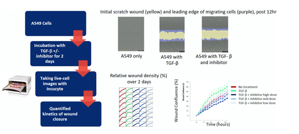
- Scratch Wound Assay with Incucyte S3 Live-Cell Analysis System
- Precision Cut Lung Slice Assay
Scratch Wound Assay